Case:
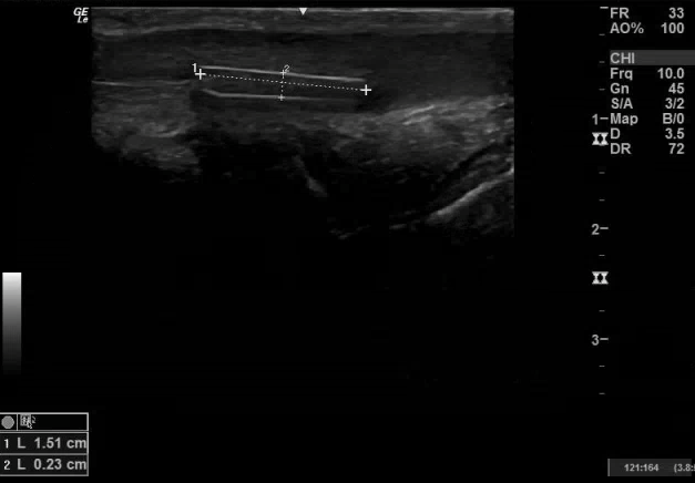
57 yo male with PMH of schizoaffective disorder presented from an inpatient psychiatric unit to the ED complaining of a foreign body (FB) in his urethra. He has a history of inserting FBs into his ears. The patient reported that he inserted a plastic fork handle into his urethra about one week ago and was now straining to void, although still passing urine. On physical exam, there was a FB palpable under the skin on the ventral side of the penis. Point-of-care ultrasound (POCUS) was performed:

Case conclusion:
After the presence of an FB was confirmed on POCUS, urology was consulted and was able to directly visualize and extract two plastic shards from the distal urethra. Unsure if there was still a deeper retained FB, urology then asked for a repeat POCUS, which demonstrated one more FB in the more proximal urethra:
Urology subsequently performed a cystoscopy and removed one last plastic shard from the urethra:

Ultrasound and Foreign Bodies:
Studies have found that ~ 38% of FBs are missed on initial physical exam. Although glass FBs will be visualized on XR in 85% of cases, other materials that are more radiolucent will often not appear on radiography. Wood, for example, is seen on XR less than 15% of the time. Studies have found that ultrasound performs very well in visualizing multiple types of FBs, including metal, glass, wood, and plastic. For the visualization of an FB in superficial body tissue and muscles, ultrasound has been found in some studies to perform better than XR or CT. Ultrasound is particularly valuable in the evaluation of FBs with low radiopacity (wood, plastic).
The surfaces of FBs always appear hyperechoic (bright) on ultrasound. If the FB has been retained for a longer time (>24 hr), surrounding inflammation can develop and appears as a hypoechoic (darker) ring around the FB. Depending on the material of the FB, it may create artifacts such as posterior shadowing or reverberation. When looking for an FB in a finger or toe, utilize a water bath for better visualization. Open wounds can be covered with a Tegaderm. Ultrasound can also be used to actively guide foreign body retrieval with in-plane needling.
Complications of missed FBs include infection and, more rarely, nerve injury. Consider POCUS as the initial screening modality when evaluating for a retained FB.
Pitfalls
Deeper FBs or those close to bone are often not well-visualized on ultrasound. False positives can occur as air, calcifications, and scar tissues may appear similar.
- Jessica Patterson, MD, Emergency Medicine Ultrasound Fellow
References:
1.https://www.ncbi.nlm.nih.gov/pmc/articles/PMC3520196/
2.https://www.ncbi.nlm.nih.gov/pmc/articles/PMC4983782/
3. http://www.emdocs.net/ultrasound-for-retained-radiolucent-foreign-body-in-soft-tissue/