Regional anesthesia is the approach of providing pain relief or anesthesia to a targeted region of the body, as opposed to a general approach which utilizes systemic medications, or via a local approach in which an anesthetic agent is injected directly into the desired site (i.e. a laceration). Regional anesthesia can be achieved in the ED via ultrasound-guided nerve blocks (UGNB). UGNBs are a valuable tool in the kit of ED pain management and procedural skills.
Via an UGNB, the provider can hijack the pain receptors "mid-stream" between the central nervous system and the area of desired anesthesia. Thus, the provider can achieve a greater general area of anesthesia (compared to local), anesthetize areas that are difficult to locally reach (i.e. a hip joint), avoid the use of systemic medications with significant side effect profiles/toxicity, and avoid injecting local anesthetic into a painful site (abscess) or where it may cause cosmetic distortion of the wound. And best of all - they are relatively safe, a fun and quick procedure to perform, and patients love them!
In order to tackle UGNBs, it's important to first understand a few key concepts:
Anatomy of Peripheral Nerves
In the periphery of the body, nerves travel within fascia "layers" or planes. These fascial planes typically exist between muscles or between a muscle and soft tissue. As the muscles in the periphery contract and relax, peripheral nerves are protected by their fascial layers which "glide" between muscles.
We utilize these fascial planes to deliver anesthetic to a nerve. By injecting anesthetic within a fascial plane near a nerve, the anesthetic will passively spread around the nerve and will diffuse across the protective sheaths around the nerve bundles (epineurium and perineurium layers) and then will act on receptors on the nerve axons. The goal is always to insert the needle tip into the space between the fascial layers, never into the nerve itself, which can cause edema and swelling within the nerve and potentially lead to neuron necrosis.


Once the anesthetic is injected, you should see "hydrodissection" of the fascial plane. The top bright white echogenic fascial layer will "lift up" as the fluid accumulates between the layers, creating a potential space. The fluid will then encircle the nerve, sometimes only in a partial or "crescent" shape. That's ok though - once you see fluid either partially or completely around the nerve, the block is going to work.

Probe choice
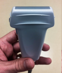
All nerve blocks should be done with a high-frequency linear probe for optimal visualization of the nerve and surrounding structures.
Technique
UGNBs can be performed utilizing an "out-of plane" or "in-plane" approach. We recommend using the "in-plane" technique for most UGNBs so that your needle tip is always in view, which theoretically minimizes the risk of passing your target and ending up with your needle tip in a neuron fascicle or nearby artery. While this technique can initially seem difficult to master, as the probe must be carefully aligned with the needle and even small probe movements can result in temporary loss of visualization of the needle, with a little practice and experience it quickly becomes easier.
References
•http://highlandultrasound.com
•Tintinalli’s 7th Ed
•www.asra.com