Case:
A 52-year-old male with PMH of HTN, IDDM presented with sudden onset of vision loss in his left eye x 1 day. He denied trauma or any inciting event. He reported that he could now only see “shadows” from his left eye. He denied photophobia, headache, fever/chills. Physical exam of the external eye was unremarkable. Visual acuity in the right eye was 20/50; the patient was unable to visualize the chart with his left. A POCUS was performed and showed the following:

Case Conclusion:
The POCUS images are consistent with both a vitreous hemorrhage and retinal detachment. Ophthalmology was consulted and confirmed the diagnoses: a retinal detachment, which they suspected was chronic, in addition to a large vitreous hemorrhage that was likely a complication of diabetic retinopathy.
Retinal Detachment vs. Vitreous Hemorrhage: Ocular POCUS
Emergency bedside ultrasound has been found to be both highly specific and sensitive for ocular pathology. Two conditions that can be easily visualized on POCUS are retinal detachment and vitreous hemorrhage.
A quick reminder on the setup for ocular ultrasound: have the patient in a reclined position and cover the closed eye with a clear adhesive dressing such as Tegaderm (you can put ointment on the eyelashes first to help the "seal"). Make sure there are no air pockets under the adhesive as this will interfere with your image. Cover the protected eye generously with gel - the more gel that you use, the less pressure you will need to apply with the probe.
Retinal detachments appear as an echogenic (bright white) ribbon-like membrane that appears to undulate or float in the back of the posterior chamber. You can confirm a retinal detachment by visualizing the membrane tethering to the optic disc (which is how it can be differentiated from a vitreous hemorrhage or detachment, in which case the mobile echogenic material crosses the optic disc). Retinal detachment example:

Retinal Detachment Tethering at Optic Disc
Vitreous hemorrhages appear as debris-like, mildly echogenic material in the posterior chamber that is mobile with eye movement. When the patient looks side to side during the ultrasound, the vitreous hemorrhage will swirl and move - its appearance has been described as looking like clothes tumbling in a drier. Vitreous hemorrhages can be missed if the gain is too low - to correctly set the gain to look for a vitreous hemorrhage, turn it up until you see artifact in the entire posterior chamber. Then, slowly lower the gain until the artifact disappears. Retinal detachments, in contrast, are typically easily visualized at "normal" gain. The echogenic material in a vitreous hemorrhage is not tethered to the optic disc. Vitreous hemorrhage example:
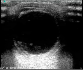
Vitreous hemorrhage
Of course, to make things confusing, these two conditions can occur simultaneously as demonstrated in the case this week.
Pearls
Ask the patient to look side to side to better visualize any echogenic material in the posterior chamber.
If you see a ribbon-like membrane, carefully evaluate it (in both the transverse and sagittal planes) to see if it tethers to the optic disc, thus confirming the presence of a retinal detachment.
Make sure your gain is set high enough to visualize a vitreous hemorrhage.
- Jessica Patterson, MD, Ultrasound Fellow
References
1. https://www.aliem.com/2014/03/ocular-ultrasound-retinal-detachment-posterior-vitreous-detachment/
2. https://www.ultrasoundoftheweek.com/uotw-12-answer/
3. Yoonessi R, Hussain A, Jang TB. Bedside Ocular Ultrasound for the Detection of Retinal Detachment in the Emergency Department. Acad Emerg Med. 2010;17(9):913–917. PMID 20836770
4. Blaivas M, Theodoro D, Sierzenski PR. A study of bedside ocular ultrasonography in the emergency department. Acad Emerg Med. 2002 Aug;9(8):791-9