• About
  • Good Times, Good Friends!
  • AEMUS & IM POCUS Fellowships
  • Faculty
  • Lewis Katz School of Medicine
  • Pocket Guide
  • Lectures
  • Tips & Tricks
  • Protocols
  • Case Challenges
  • Journal Club
  • Regional Anesthesia
  • Research
  • Upcoming Events
  • Philadelphia POCUS Conference
  • Breaking News
  • SonoPatch
  • Contact
  • Philly UltraFest
  • Search
Menu

Temple Point-of-Care Ultrasound

1316 West Ontario Street
Philadelphia, PA, 19140
GMAIL - TempleHEALTHPOCUS

Your Custom Text Here

Temple Point-of-Care Ultrasound

  • About
  • Good Times, Good Friends!
  • AEMUS & IM POCUS Fellowships
  • Faculty
  • Lewis Katz School of Medicine
  • Pocket Guide
  • Lectures
  • Tips & Tricks
  • Protocols
  • Case Challenges
  • Journal Club
  • Regional Anesthesia
  • Research
  • Upcoming Events
  • Philadelphia POCUS Conference
  • Breaking News
  • SonoPatch
  • Contact
  • Philly UltraFest
  • Search

PE Protocol

October 28, 2018 Mark Magee
PE protocol.png

1. Kearon C, Ginsberg JS, Hirsh J. The role of venous ultrasonography in the diagnosis of suspected deep venous thrombosis and pulmonary embolism. Ann Intern Med. 1998;129:1044–1049.

2. Konstantinides SV, Torbicki A, Agnelli G, Danchin N, Fitzmaurice D, Galiè N, Gibbs JS, Huisman MV, Humbert M, Kucher N, Lang I, Lankeit M, Lekakis J, Maack C, Mayer E, Meneveau N, Perrier A, Pruszczyk P, Rasmussen LH, Schindler TH, Svitil P, Vonk Noordegraaf A, Zamorano JL, Zompatori M. ESC Guidelines on the diagnosis and management of acute pulmonary embolism: the Task Force for the Diagnosis and Management of Acute Pulmonary Embolism of the European Society of Cardiology (ESC) Endorsed by the European Respiratory Society (ERS) Eur Heart J. 2014;35:3033–3073.

3. Le Gal G, Righini M, Sanchez O, Roy PM, Baba-Ahmed M, Perrier A, Bounameaux H. A positive compression ultrasonography of the lower limb veins is highly predictive of pulmonary embolism on computed tomography in suspected patients. Thromb Haemost. 2006;95:963–966.

4. Mathis G, Blank W, Reissig A, Lechleitner P, Reuss J, Schuler A, Beckh S. Thoracic ultrasound for diagnosing pulmonary embolism: a prospective multicenter study of 352 patients. Chest. 2005;128:1531–1538. [PubMed]

5. McConnell MV et al. Regional Right Ventricular Dysfunction Detected by Echocardiography in Acute Pulmonary Embolism Am J Cardiol 1996

6. Squizzato A, Rancan E, Dentali F, Bonzini M, Guasti L, Steidl L, Mathis G, Ageno W. Diagnostic accuracy of lung ultrasound for pulmonary embolism: a systematic review and meta-analysis. J Thromb Haemost. 2013;11:1269–1278.
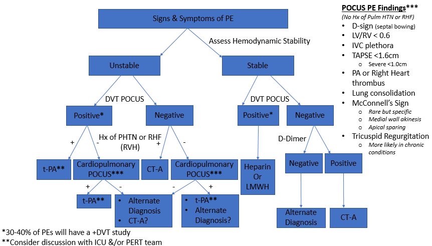
In PE Protocol
← Lung Ultrasound Protocol for COVID=19Fluid Responsiveness vs Tolerance →

Archives

Tips & Tricks
  • Cardiac Ultrasound
  • Lung Ultrasound
  • USG TLC Placement
Protocols
  • 1st Trimester Protocol
  • AAA Protocol
  • COVID-19 Protocol
  • DVT Protocol
  • Fluid Responsiveness
  • IVC Fluid Status Protocol
  • PE Protocol
  • Pericardial Eff Protocol
Case Challenges
  • Aorta Ultrasound
  • Bowel Ultrasound
  • Cardiac Ultrasound
  • DVT Ultrasound
  • HEENT ultrasound
  • Ocular Ultrasound
  • Pelvic Ultrasound
  • Pulmonary Ultrasound
  • RUQ Ultrasound
  • Renal Ultrasound
  • Soft Tissue Ultrasound
  • Testicular Ultrasound
  • Vascular Ultrasound
  • eFAST Ultrasound
Journal Club
  • Abdominal JC
  • Aorta JC
  • Biliary JC
  • Cardiac JC
  • Cardiopulmonary JC
  • Carotid Flow Time JC
  • HEENT JC
  • LUS JC
  • MSK JC
  • Ob/Gyn JC
  • Ocular JC
  • POCUS Basics JC
  • Pediatrics JC
  • Procedures JC
  • Regional Anesthesia JC
  • Renal Ultrasound JC
  • Soft Tissue JC
  • Trauma JC
  • Vascular JC
  • Volume Assess/Resusc JC
Regional Anesthesia
  • Fascia Iliaca
  • Introduction
  • Serratus Anterior

Subscribe

Sign up with your email address to receive news and updates.

We respect your privacy.

Thank you!