The Article: Prabhu, S. Kanal, K. Bhargava, P. Vaidya, S. Dighe, M. “Ultrasound artifacts: classification, applied physics with illustrations, and imaging appearances.” Ultrasound Quarterly 2014:6(30).
The Idea: The use of ultrasound has become increasingly more common, providing a safe and portable diagnostic tool. Despite advances in technology and operator training, artifacts resulting from physiology and ultrasound hardware can limit imaging and cause difficulties in interpretation. This review classifies several types of ultrasound artifacts, providing brief explanations of the physics behind the artifacts and strategies for minimizing or utilizing artifact to our advantage.
The Review: This review classifies artifacts based on if the artifact is observed in B-mode or Doppler imaging. Furthermore, within each classification, the authors break artifacts into one of two subcategories: those ‘related to structure’ and those ‘related to beam properties.’ Below is an example of each and a brief explanation of the physics as provided by the authors.
Artifact in B mode related to structures of the tissue that is being imaged
-Examples include: mirror-image artifact, shadowing artifact, comet tail artifact enhancement artifact, speckle artifact and reverberation artifact
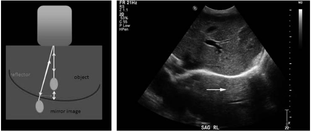
Mirror Artifact
This occurs when the plane being imaged contains an object next to a strong reflector. Reverberations of the ultrasound beam occur between the object and strong reflector, which prolongs the time of the ultrasound beam. This artifact creates a mirror image of the object on the opposite side of the reflector. Strong reflectors include the diaphragm, the pleura, the bladder and the bowel. This artifact can be troublesome when it is difficult to distinguish if the image generated is due to reflections of bowel/bladder or is true pathology.
Artifact in B mode related to the properties of the ultrasound beam and processing
-Examples include: refraction artifact, beam width artifact, range ambiguity artifact, speed error artifact
Speed Error Artifact
This artifact occurs because the ultrasound programming assumes that the velocity of sound in tissue is a constant 1540m/s. The ultrasound hardware determines a depth of an imaged object based on this velocity constant and the round-trip flight time of the sound beam. However, in fatty tissue, the speed of sound is less than 1540m/s. Thus, in tissues containing fat, the depth of the object displayed will appear deeper than its true location. Conversely, tissues with a velocity of sound greater than 1540m/s, such as skeletal muscle, objects will appear shallower than their true location. The authors suggest that this artifact can be clinically useful as it can help determine the composition of a lesion in a solid organ.

Artifact in Doppler related to properties of the tissue
-Examples include: spectral broadening artifact, vascular motion artifact
Vascular Motion Artifact
This occurs when the transducer is held still, but there is movement of the vessel due to outside forces, creating artificial flow patterns and potentially the false appearance of pulsation. This is clinically observed in the portal vein, where the motion of the heart can be transmitted to the liver and onto the portal vein. The authors suggest increasing the size of the Doppler gate to include the entire width of the vessel, and to image other portal branches.

Artifact in Doppler related to beam properties and processing
-Examples include: Aliasing artifact, direction ambiguity artifact, color flow artifact, twinkle artifact
Directional Ambiguity
This artifact is observed most often in solid organs where the vessels travel in many directions and orientations. In the case, the operator is evaluating a vessel with an ultrasound beam perpendicular to the direction of flow. The beam side lobes interpret flow as both upstream and downstream. This results in a tracing with movement above and below the baseline. This artifact can be avoided by angling the transducer away from the perpendicular.


Takeaways: Ultrasonography is an invaluable tool in the rapid assessment and diagnosis of patients in the Emergency Department. This review demonstrates numerous artifacts generated by the operator, the qualities of the tissue being imaged, and the machinery and programming of the ultrasound machine. While operators can mitigate some types of artifact, awareness of inherent and unavoidable artifact is critical in understanding the image generated, its reliability and diagnostic value.
Post by Dr. Katelyn Peloza, EM PGY 1